White patches in the oral cavity. How to detect and prevent potentially malignant changes
Oral leukoplakia is a chronic mucosal lesion that looks like a white or grey patch in the oral cavity, and usually does not cause any complaints. It is one of the most common potentially malignant diseases, affecting approximately 3.41-11.74% of the general population. However, not all leukoplakia develops into a malignant tumour.
The most common risk factors for oral leukoplakia are smoking, alcohol use and human papillomavirus infection. In recent years
in Latvia, young people are increasingly using smokeless tobacco, which may also contribute to the development of precancerous and malignant tumours.
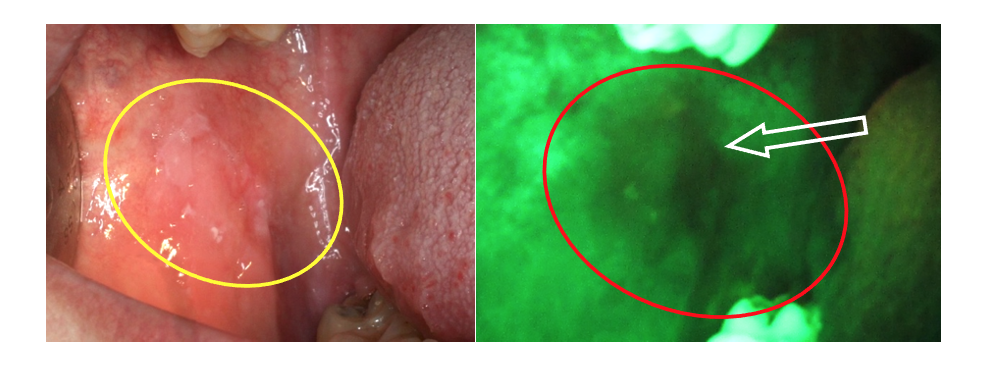
The aim of the doctoral thesis by Madara Dzudzilo was to assess clinical, morphological and immunohistochemical changes of oral cavity leukoplakia in conjunction with salivary test parameters to identify early signs of oral leukoplakia malignisation. VELscope fluorescence spectroscopy images were taken during the research, which allowed for the assessment of the borders of abnormal tissues.
 Leukoplakia in the mucosa of the cheeks under artificial light (left) and VELscope (right). The arrow shows the most visible loss of fluorescence. Photo from the archive of the Oral Medicine Centre of the Institute of Stomatology
According to the research results, PhD student Dzudzilo concluded that in their study group
oral leukoplakia was more common in men with an average age of 57 years, and the most frequent site was the mucosa of the cheeks and the back of the tongue.
It should be noted that dentists recommend that patients undergo soluble CD44 and total protein saliva tests to screen for the risk of malignant changes in leukoplakia, as well as early diagnosis of recurrent leukoplakia without surgical intervention.
Read more


