Study on genes and gene proteins in different tissues affected by facial clefts
Facial clefts are a congenital development defect caused by the defective fusion of facial folds before birth. Clefts can be unilateral or bilateral and can affect the oral mucosal tissues, forming clefts of the lip and palate.
The formation of facial clefts has been linked to cleft genes, whose proteins regulate the growth and formation of tissues in the facial region, but the involvement of these genes in the formation of facial clefts is relatively unclear and has not been particularly studied in human tissues.
The aim of the thesis was to determine the distribution and relative number of genes and proteins in different types of cleft-affected tissue.
This information could potentially provide a better understanding of cleft formation in humans and could lead to improvements in the treatment and prevention of clefts in the future.
This study assessed the relative number of immunopositive structures of BarH-like homeobox 1 (BARX1), distal-less homeobox 4 (DLX4), forkhead box E1 (FOXE1), homeobox B3 (HOXB3), muscle segment homeobox 2 (MSX2), paired box transcription factor 7 (PAX7), paired box transcription factor 9 (PAX9), receptor-like tyrosine kinase (RYK), Sonic hedgehog (SHH), SRY-box transcription factor 3 (SOX3), wingless-type MMTV integration site protein 3A (WNT3A) and wingless-type MMTV integration site protein 9B (WNT9B) using immunohistochemistry method. These cleft gene-derived proteins regulate certain aspects of facial region development not only in clefts but also in healthy oral mucosal tissues, either by promoting, inhibiting, or modifying epithelial and connective tissue growth. These gene disorders are associated with the development of facial clefts in humans.
The results of the study showed that healthy oral tissues had the highest number of cells containing WNT9B, WNT3A, SOX3 cells, moderate numbers of SHH, PAX7, HOXB3, FOXE1, lower numbers of PAX9, DLX4, RYK, but no BARX1 and MSX2. In contrast, cleft-affected tissues showed an increase in BARX1 and MSX2 and a decrease in SOX3, WNT3A and WNT9B. Unilateral cleft is characterised by a more pronounced finding of BARX1, FOXE1, HOXB3 and PAX7, indicating an increase in cell renewal potential. Bilateral cleft is considered a more clinically severe type of cleft, and it is characterised by an increase in DLX4 and PAX9 and a decrease in SHH, suggesting a functional role of these factors specifically in tissues affected by bilateral cleft. The study showed that all types of cleft (unilateral, bilateral and cleft palate) were characterised by an increase in MSX2 and RYK, while reductions were observed for SOX3, WNT3A and WNT9B in the tissue.
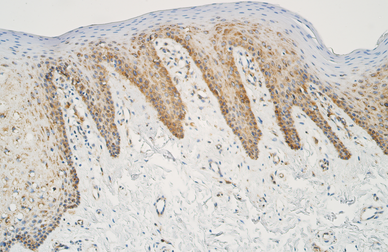
Moderate to numerous Sonic hedgehog (SHH) positive epithelial cells in a patient with a complete bilateral cleft lip and palate. The tissues were obtained during lip surgery. SHH immunohistochemistry (× 200). Image from the thesis by Mārtiņš Vaivads
Mārtiņš Vaivads will defend his doctoral thesis Investigation of Genes and Gene Proteins in Cleft Affected Tissue on 14 December 2023. Read more
Related news
 Time capsule laid at ceremony for construction of new RSU sports complexFor Students, Consolidation, For RSU Employees, Internal consolidation, Development