How do cholesteatomas, benign but locally aggressive formations in the middle ear, develop?
Images from Kristaps Dambergs' doctoral thesis
Cholesteatomas can be congenital or acquired. Acquired cholesteatomas occur when skin cells (epithelium) lodge in the middle ear, where they should not be, and transform into cholesteatoma tissue.
Cholesteatoma tissue is hyperproliferative, meaning that the cells grow and multiply excessively compared to normal cell growth, and they degrade the surrounding tissue in the middle ear.
Although the formation is rare, with an average incidence in Europe of 7 per 100,000 people a year, treating it completely is difficult. Recurrences are often expected.
The most common complaints with cholesteatomas are loss of hearing and otorrhea that recur frequently. Cholesteatomas can also cause complications such as meningitis, brain abscesses, facial nerve paralysis and sigmoid sinus thrombosis.
Cholesteatomas develop as part of a complicated process where the disease “transforms” surrounding normal tissue into damaged. This process leads to uncontrolled tissue growth and the transformation of surrounding tissues, resulting in inflammation.
The aim of the PhD thesis by Kristaps Dambergs, a doctoral student at Rīga Stradiņš University (RSU) and an assistant at the RSU Department of Otorhinolaryngology, was to determine and describe how cholesteatomas develop, what changes they cause in tissues, and what the interaction of the new formation is in tissues of patients of different ages.
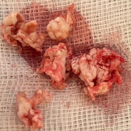
 Tissue sample of acquired cholesteatoma. Surgery material
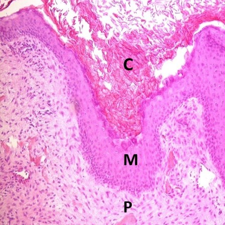
 Adult cholesteatoma. C (cystic layer); M (matrix); P (perimatrix). Haematoxylin and eosin, × 200
Until now, selective tissue changes, such as remodelling, proliferation, inflammation and local tissue defence, have been studied sporadically and unrelated in patients with cholesteatoma. This study examines these tissue changes comprehensively. The study selects twelve different tissue factors that characterise the clinical symptoms of cholesteatoma and which is the largest number of tissue factors used in a single study of children and adult cholesteatoma.
Each factor was examined and described, analysing a total of more than 700 micro-preparations.
The study included cholesteatoma tissue from 50 patients. The patients were divided into two age groups - children and adults. Each group consisted of 25 patients. The control group consisted of the skin of external ear meatus, which was obtained from the bodies of 7 deceased people.
Read more


